

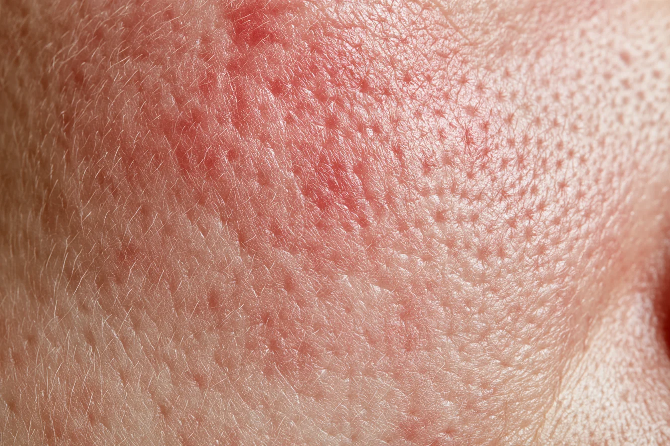
We often hear this in clinic: "My skin got even redder and broke out after scar laser." "My face is red to begin with. Can I still have scar treatment?" When scars are your concern, it is natural to look only at the scars, and it can seem that the stronger the device, the faster things will improve. Yet two people can have the same treatment and one improves while the other gets redder. Why are scars on a red base harder to treat, and why do some skins get worse after scar treatment? This column follows that one question through to the end, through the mechanisms inside the skin and the research.
Summary in Three Lines
- Scar treatment deliberately sets small, controlled fires so the skin rebuilds. When the base skin is already red with dilated vessels, it can be shaken more by the same stimulation.
- That does not mean red skin cannot have scar treatment. The issue is not the stimulation itself but its intensity, order and purpose. There is even a study in which redness and inflammation from rosacea decreased after microneedle RF.
- So we settle the underlying inflammation and vascular reactivity first, or alongside scar treatment. Once the base is calmer, the scar can receive the treatment it actually needs.
The Short Answer — The Ground Under the Scar Is Different
Put simply, scar treatment deliberately sets small fires so the skin rebuilds itself. On damp ground, a small fire burns only as far as planned and goes out. On bone-dry ground, the same fire can spread sideways. The point of this column is that a base that is already red, with dilated vessels, resembles that dry ground.
When the base skin is already red with dilated vessels, it can be shaken more by the new stimulation of scar treatment. That does not mean red skin cannot have scar treatment. The issue is not the stimulation itself but its intensity, order and purpose.
In fact, a randomized study of red acne scars reported differences in both the scars and the redness between adding a vascular laser to fractional laser in the same session and not adding it (Zhou et al., 2024), and in rosacea skin, redness and inflammation actually decreased after microneedle RF (Park et al., 2016). That is why we settle the base first, or together with scar treatment. Dampening the ground first lets us put in the fire the scar actually needs.
First, Separate What "Red" Means — Four Kinds of Redness
The single word "red" covers four quite different things.
- Redness from acne marks (PIE) — dot-like redness left where acne has passed
- Redness of the scar itself — a depressed scar that is red
- Redness of the base — the whole face is red, regardless of where acne has been
- Redness after a procedure — a reaction that appears after treatment
This column is about the third.
The first kind, PIE, is a trace where a little of the inflammatory ember remains underneath even though the surface has settled. That is why we explain to acne patients that the sooner these red marks are dealt with, the less scarring tends to form, and we treat them actively with vascular laser. We cover PIE in detail in our column on post-inflammatory erythema (PIE). The second kind, redness of the scar itself, also often responds well to microneedle RF or vascular laser. Lumping all redness together can send treatment in the wrong direction.
Base redness is not one thing either
- Flushing — the face flares up with heat or irritation and then settles
- Rosacea — redness becomes fixed, often with burning or papules
- Telangiectasia — dilated, visible small vessels
- Sensitive skin — everything stings and the skin flares easily
Most people have a mix, but they are not the same thing. The tissue studies below are rosacea studies, so they cannot simply be applied to someone with a few visible vessels or sensitive skin. We discuss how to sort types of redness in detail in our column on V-Beam for diffuse redness.
A Rosacea Base Is More Than Dilated Vessels — Skin Whose Alarm Goes Off Easily
Let us translate "dry ground" into the language of the skin. Rosacea skin is not just skin with dilated vessels; its immune environment is somewhat different. The alarm goes off at small stimuli, and once it goes off, there is a loop that readily spreads it into inflammation.
The skin has sensors that detect danger signals (TLR2). In a study comparing 11 people with rosacea and 8 with normal skin, these sensors were more abundant in rosacea, and when stimulated they released more of a protein-cutting enzyme (KLK5) (Yamasaki et al., 2011, human tissue, cell and mouse experiments). This enzyme turns the skin's antimicrobial substance LL-37 into fragments that cause inflammation (Yamasaki et al., 2007, human tissue and mouse experiments).
Mast cells (immune cells involved in allergic reactions) join in as well. Comparing the skin of 6 people with rosacea and 6 healthy people, mast cell-related enzymes were increased, and in mouse experiments, without mast cells, injecting LL-37 did not produce rosacea-like inflammation (Muto et al., 2014).
No papules on the surface does not mean no inflammation. In a retrospective analysis of tissue from 226 people, clusters of inflammatory cells around vessels and follicles were common in both the redness-only type and the papular type (Lee et al., 2016, retrospective tissue study). In a study of gene expression in facial tissue, inflammatory cells were already present even in the redness-only type, with increased macrophages and mast cells (Buhl et al., 2015, human tissue study).
The burning has a reason too. Channels that respond to heat and other triggers (TRPV) were increased in redness-only rosacea skin (Sulk et al., 2012, human tissue study), and sensory nerves were reported lying right next to blood vessels and mast cells (Schwab et al., 2011, human tissue study). It is skin in which vessels, immune cells and nerves sit close together and react together.
Imagine placing a heat-based treatment that is meant to make the skin rebuild on top of this, and you can see why we become cautious.
How Scars Form on a Red Base — Where Lingering Inflammation Meets Collagen Breakdown
So how does this base connect to scar formation? When acne in people prone to scarring is examined in tissue, the vascular reaction does not settle even during recovery, and inflammatory cells linger late (Holland et al., 2004; Carlavan et al., 2018, tissue studies of back lesions). Set side by side with a rosacea base, the two resemble each other in three places.
How long inflammation lingers
In people prone to scarring, acne inflammation lasts longer, while in rosacea skin, inflammatory cells remain around vessels and follicles even when there is no acne. It is as though the surroundings are already heated before the ember of acne inflammation, the inflammation left underneath even when it seems to have gone out, has even settled in.
Enzymes that cut collagen
Acne tissue showed more collagen-cutting enzymes (MMPs) and more broken collagen (Kang et al., 2005, facial tissue study), and rosacea skin also showed increased MMP-9 from the same family (Muto et al., 2014). The same paper also reported that mast cells stimulated by LL-37 release this enzyme.
Blood vessels and lymphatics
When a wound heals, dilated vessels should return to normal, but in poorly healing wounds and scars this process is reported to go awry (Han et al., 2022, wound repair review). Rosacea skin starts with more vessels and lymphatics (Schwab et al., 2011; Gomaa et al., 2007, human tissue studies), so it may be a base with more to sort out.
So we see a red base as possibly being dry ground where acne inflammation can linger longer. To be clear, we did not find a study that directly compares acne scar formation in people with and without rosacea. What we can say is that when scar research and rosacea research are placed side by side, they point in a similar direction.
There is also evidence pointing the other way. Long-standing rosacea can lead to thickening (fibrosis) rather than tissue loss (Steinhoff et al., 2011, review), and LL-37 is not only an inflammatory substance; it is also used in normal wound healing (Heilborn et al., 2003, human skin study). Nor is there a figure for how much collagen is lost in rosacea skin. So this does not mean rosacea skin cannot regenerate. Please read it only as an environment where the same events may drag on longer.
When Sensitive Skin Gets Worse After Scar Treatment — Treatment Is a Small Fire Too
Back to the opening words: "My skin got even redder and broke out after scar laser." Scar treatment deliberately creates small injuries: fractional laser with heat, microneedle RF with needles and radiofrequency, and subcision by cutting tethering with a needle. In skin with a stable base, that fire burns as planned and goes out, and regeneration begins. If the ground is dry, the same fire can spread.
What we usually see in sensitive skin after scar treatment falls along these lines:
- Flushing or rosacea flares up
- Redness lasts a long time
- Pigmentation is left behind
- Contact dermatitis or acne-like rashes appear
How far does the literature go? There is a case of a 61-year-old woman who developed rosacea symptoms after microneedle RF for wrinkles (Aşiran Serdar et al., 2019, single case), and a case in which rosacea worsened after a collagen stimulator (PLLA) was injected with a microneedle device (Yu et al., 2022, single case). Neither involved scar treatment, and each is a single case. The microneedle RF case conflicts with studies in which the same type of device improved rosacea, and its authors point that out themselves. There is also a systematic review of complications of facial procedures in people with skin conditions, but most of the evidence comes from case reports and small case series, so it cannot tell us how often this happens (Perez et al., 2026).
There is also a reason not to brush off redness after a procedure. In a randomized study treating 67 solar lentigines in 28 people with a 532 nm pigment laser, increased redness at 2 weeks was a risk factor for hyperpigmentation (Kang et al., 2024, odds ratio 1.32). This is a pigment laser study, not a scar procedure, and it looked at redness that increased after treatment rather than redness that was there beforehand, so it is only indirect evidence for scar treatment. We cover pigmentation in our column on post-inflammatory hyperpigmentation.
Swelling can be another way it shows up. One author reported seeing cases of facial swelling lasting more than 6 months after extensive, aggressive subcision (Lim, 2025). The author speculates that when lymphatic drainage is weakened in skin with long-standing inflammation, wide undermining may increase swelling. It is an opinion based on case experience, and the author states plainly that objective data are lacking.
So how much more likely is a flare with rosacea? There is no number. We did not find a study comparing flare rates after scar procedures by whether rosacea was present, nor one that defined sensitive skin separately and compared recovery times. That is why, on this point, we can only speak from clinical experience rather than published data.
You may also have heard that the longer the redness lasts after a procedure, the better the collagen is being built. We have not found evidence to support that. Redness that does not improve and lingers, or that comes with burning or a rash, is redness that needs to be checked. The treatment order for skin with a weak barrier is covered separately in our column on skin where pores should wait their turn.
Why "All Stimulation Is Bad" Is Only Half Right — Microneedle RF and Vascular Laser Studies
Should red skin, then, avoid needles and lasers altogether? No. Saying that fire can spread on dry ground does not mean fire should never be used. In fact, there are studies pointing the other way.
In a randomized study of 21 people with mild to moderate rosacea, one cheek received microneedle RF twice, 4 weeks apart, and the other cheek was left untreated (Park et al., 2016). The treated side improved clinically in 17 people (81%), and the erythema index fell by 13.6% at 12 weeks. In tissue, dermal inflammation and mast cells also decreased. It is interesting that the mast cells caught up in the alarm loop decreased after a designed stimulus. Still, the authors themselves described the improvement as modest, and this was a small trial designed to treat rosacea, not scars.
In a retrospective study of 25 people with papulopustular rosacea that had not responded well to medication, who received three sessions of vascular laser (PDL) and microneedle RF while taking low-dose isotretinoin (Kwon et al., 2020), papules decreased by 71% and the erythema index by 54% at 16 weeks after the last treatment. It had no comparison group, and because medication was used together, the result should not be read as the effect of the laser alone.
There is a study of red acne scars as well. Ninety people were randomly assigned to non-ablative fractional laser alone or with a vascular laser (595 nm PDL) added in the same session (Zhou et al., 2024). At 4 weeks, the scar score (lower is better) was 6.67 with non-ablative fractional alone and 4.98 with the combination, and the proportion with reduced redness was 66.7% and 88.9%. It was a single-session study with 4 weeks of follow-up, and 88.9% is the proportion whose redness decreased. Color change may also have been reflected in the scar score. Non-ablative fractional laser is not a device we have at our clinic, so we mention it only as part of the study design.
There is a similar story in skin with active acne. In a randomized split-face trial of 23 Koreans with both acne and scars, in which one half of the face received microneedle RF plus fractional CO₂ and the other half fractional CO₂ alone (Kim et al., 2023), inflammatory acne decreased by 46.3% and 12.0% at 20 weeks. This was an active acne study, so it cannot be applied directly to skin with rosacea or flushing, and the devices differ between studies, so they are not necessarily the same models as ours.
It is in the same vein that we noted in our Potenza scar column that immature scars still carrying redness sometimes respond well to microneedle RF.
Why does the same microneedle RF help some people and cause flares in others?
Here is the answer: the issue is not the stimulation itself but its intensity, order and purpose. Just as Potenza for pores and Potenza for scars are not the same treatment, the same device is used at one intensity to treat scars and another to calm the base (Same Device, Different Results; Why Does Potenza Need a Design?). The same fire becomes something entirely different depending on where it is lit, how much and why.
Dampening the Base First — How We Look at Scars on Red Skin
So when we look at scars on a red base, we do not start by measuring the depressions. There are things we ask first.
- Is your face red even on ordinary days?
- Does it burn or sting?
- Have you ever used a steroid ointment on your face?
- Are new acne spots still appearing?
- How long did redness last after previous procedures?
These are clues to how dry the ground is. The first two tell us the current state of the base, the fourth tells us about the fire burning now, and the fifth tells us how this skin reacted to the last fire. You will see why the third is on the list in our column on rosacea misdiagnosis.
If the base is dry, we build into the plan the work of dampening it, before scar treatment or alongside it. Dampening here means calming the inflammation and vascular reactivity. Sometimes medication is needed; sometimes vascular laser or LDM is the right fit.
That does not mean treating the scars gently forever. Going too gently tends to end in "I had it done, but it did not do much." The reason for dampening the base is precisely to make ground on which the scar can receive the fire it needs. Preparing the ground first is one step back for two steps forward.
Putting the red acne scar study above (Zhou et al., 2024) together with the study in which erythema, dermal inflammation and mast cells decreased after microneedle RF in rosacea skin (Park et al., 2016), we believe that settling the vascular and inflammatory reactions of the base helps scar treatment. Whether to settle it first or address it within the same plan should be decided after looking at the state of the base.
In Short — Treating the Base Along with the Scar
Scars on a red base are hard to treat not just because vessels are dilated, but because it is ground where the alarm goes off easily and inflammation can linger. When skin gets worse after scar treatment, it may be less that the treatment was bad and more that a fire unsuited to that ground spread. This part reflects our clinical judgment.
Scar treatment means treating not only the depression but also the base it sits on. If you were upset that your skin got redder after scar treatment, or have hesitated because your face is red, we would like to look at the base together with you before you give up.
See also: telling red marks apart from depressed scars
Precautions and Limits of the Evidence
Responses to scar treatment and the course of recovery differ from person to person depending on scar shape, the condition of the base skin and previous treatment. The order and intensity of treatment are decided at consultation.
Scar treatments (fractional laser, microneedle RF, subcision) can cause redness, swelling, bruising, crusting and pigmentation, and rarely scarring. Vascular laser can cause swelling and bruising. Flushing or rosacea may worsen after a procedure. Procedures on areas with infection or severe inflammation are postponed or adjusted.
The studies in this column are a mix of tissue studies, small randomized trials, retrospective studies, case reports and author opinion, with different subjects and measurement methods, so their numbers cannot be compared directly. Tissue findings from rosacea cannot be applied to simple telangiectasia or to all sensitive skin. The effect of base flushing or rosacea on scar formation and treatment response, and the order of base treatment and scar treatment (first or together), are judgments based on mechanistic research, small clinical studies and clinical experience together, and more direct comparative research is needed. This summary is based on research confirmed to date and does not replace individual care.
References
Show 23 references
- Yamasaki K, Kanada K, Macleod DT, et al. TLR2 expression is increased in rosacea and stimulates enhanced serine protease production by keratinocytes. J Invest Dermatol. 2011;131(3):688-697. doi:10.1038/jid.2010.351
- Yamasaki K, Di Nardo A, Bardan A, et al. Increased serine protease activity and cathelicidin promotes skin inflammation in rosacea. Nat Med. 2007;13(8):975-980. doi:10.1038/nm1616
- Muto Y, Wang Z, Vanderberghe M, et al. Mast cells are key mediators of cathelicidin-initiated skin inflammation in rosacea. J Invest Dermatol. 2014;134(11):2728-2736. doi:10.1038/jid.2014.222
- Lee WJ, Jung JM, Lee YJ, et al. Histopathological analysis of 226 patients with rosacea according to rosacea subtype and severity. Am J Dermatopathol. 2016;38(5):347-352. doi:10.1097/DAD.0000000000000454
- Buhl T, Sulk M, Nowak P, et al. Molecular and morphological characterization of inflammatory infiltrate in rosacea reveals activation of Th1/Th17 pathways. J Invest Dermatol. 2015;135(9):2198-2208. doi:10.1038/jid.2015.141
- Sulk M, Seeliger S, Aubert J, et al. Distribution and expression of non-neuronal transient receptor potential (TRPV) ion channels in rosacea. J Invest Dermatol. 2012;132(4):1253-1262. doi:10.1038/jid.2011.424
- Schwab VD, Sulk M, Seeliger S, et al. Neurovascular and neuroimmune aspects in the pathophysiology of rosacea. J Investig Dermatol Symp Proc. 2011;15(1):53-62. doi:10.1038/jidsymp.2011.6
- Gomaa AH, Yaar M, Eyada MM, Bhawan J. Lymphangiogenesis and angiogenesis in non-phymatous rosacea. J Cutan Pathol. 2007;34(10):748-753. doi:10.1111/j.1600-0560.2006.00695.x
- Holland DB, Jeremy AH, Roberts SG, et al. Inflammation in acne scarring: a comparison of the responses in lesions from patients prone and not prone to scar. Br J Dermatol. 2004;150(1):72-81. doi:10.1111/j.1365-2133.2004.05749.x
- Carlavan I, Bertino B, Rivier M, et al. Atrophic scar formation in patients with acne involves long-acting immune responses with plasma cells and alteration of sebaceous glands. Br J Dermatol. 2018;179(4):906-917. doi:10.1111/bjd.16680
- Kang S, Cho S, Chung JH, et al. Inflammation and extracellular matrix degradation mediated by activated transcription factors nuclear factor-kappaB and activator protein-1 in inflammatory acne lesions in vivo. Am J Pathol. 2005;166(6):1691-1699. doi:10.1016/S0002-9440(10)62479-0
- Han C, Barakat M, DiPietro LA. Angiogenesis in wound repair: too much of a good thing? Cold Spring Harb Perspect Biol. 2022;14(10):a041225. doi:10.1101/cshperspect.a041225
- Steinhoff M, Buddenkotte J, Aubert J, et al. Clinical, cellular, and molecular aspects in the pathophysiology of rosacea. J Investig Dermatol Symp Proc. 2011;15(1):2-11. doi:10.1038/jidsymp.2011.7
- Heilborn JD, Nilsson MF, Kratz G, et al. The cathelicidin anti-microbial peptide LL-37 is involved in re-epithelialization of human skin wounds and is lacking in chronic ulcer epithelium. J Invest Dermatol. 2003;120(3):379-389. doi:10.1046/j.1523-1747.2003.12069.x
- Aşiran Serdar Z, Aktaş Karabay E. A case of fractional microneedling radiofrequency induced rosacea. J Cosmet Laser Ther. 2019;21(6):349-351. doi:10.1080/14764172.2019.1661487
- Yu S, Gong R, Wang L. Rosacea aggravated after intracutaneous injection of poly-L-lactic acid through a stamp-type microneedle. Plast Reconstr Surg Glob Open. 2022;10(11):e4655. doi:10.1097/GOX.0000000000004655
- Perez SM, Kennedy DJ, Ascher B, et al. Implications of dermatologic disorders in facial cosmetic surgery: a systematic review. Ann Plast Surg. 2026;96(5):404-412. doi:10.1097/SAP.0000000000004731
- Kang DH, Choi SM, Lee Y, et al. Postoperative risk assessment of post-inflammatory hyperpigmentation and the efficacy of delayed prevention following 532 nm Q-switched Nd:YAG laser treatment of solar lentigines: a randomized controlled study. J Dermatolog Treat. 2024;35(1):2398768. doi:10.1080/09546634.2024.2398768
- Lim DS. Subcision — the art of controlled aggression. J Cosmet Dermatol. 2025;24(1):e16746. doi:10.1111/jocd.16746
- Park SY, Kwon HH, Yoon JY, et al. Clinical and histologic effects of fractional microneedling radiofrequency treatment on rosacea. Dermatol Surg. 2016;42(12):1362-1369. doi:10.1097/DSS.0000000000000888
- Kwon HH, Jung JY, Lee WY, et al. Combined treatment of recalcitrant papulopustular rosacea involving pulsed dye laser and fractional microneedling radiofrequency with low-dose isotretinoin. J Cosmet Dermatol. 2020;19(1):105-111. doi:10.1111/jocd.12982
- Zhou C, Yao M, Chen W, et al. Treatment of erythematous acne scars using 595-nm pulsed dye laser combined with 1565-nm (…) nonablative fractional laser. J Cosmet Dermatol. 2024;23(6):2015-2021. doi:10.1111/jocd.16235
- Kim J, Lee SG, Choi S, et al. Combination of fractional microneedling radiofrequency and ablative fractional laser versus ablative fractional laser alone for acne and acne scars. Yonsei Med J. 2023;64(12):721-729. doi:10.3349/ymj.2023.0234
This column is adapted from Dr. Jeong's blog post (2026-11-10, in Korean). Read the original (Korean)
From Consultation Through Treatment
A board-certified dermatologist examines you directly, identifies which layer the cause sits in, sets the device and the parameters, and then carries out the procedure as the same physician.
The number of sessions, the intervals, the maintenance point and the cost are agreed together before the first session.
Frequently Asked Questions
- If I have flushing, can I still have scar treatment?
- Yes. It is less that red skin rules treatment out and more a matter of how unstable the base is right now, and in what order and at what intensity to proceed. If flushing or rosacea is flaring, the treatment plan can be adjusted.
- Does scar treatment work better if I have vascular laser first?
- For people with a red, sensitive base, we often plan to settle the base with vascular laser first, or alongside scar treatment. In red acne scars, a randomized study also reported differences in scar score and redness between adding vascular laser in the same session and not adding it (Zhou et al., 2024). Whether to do it first or together is decided by the state of the base, and people with a stable base sometimes go straight to scar treatment.
- How long should I wait and watch redness after a scar procedure?
- A few days of redness is a common reaction. But if it does not improve and lingers, or if burning or a rash comes with it, it is a good idea to be seen before your next treatment. Because the duration depends on the type and intensity of the procedure, it is hard to give a single number.
- I still get breakouts. Can I have scar treatment?
- There is no rule that you must be completely free of acne to begin. If inflammation is burning strongly, we suggest putting it out first; if only a little remains, we may address it within the same treatment plan. This decision, too, is made in clinic after looking at your skin.
This article is general information and does not replace an individual diagnosis or treatment. Because the right decision depends on your symptoms and skin condition, please see a board-certified dermatologist.