

Medically reviewed by Dr. Jeong Kyungmuk, board-certified dermatologist · Last updated
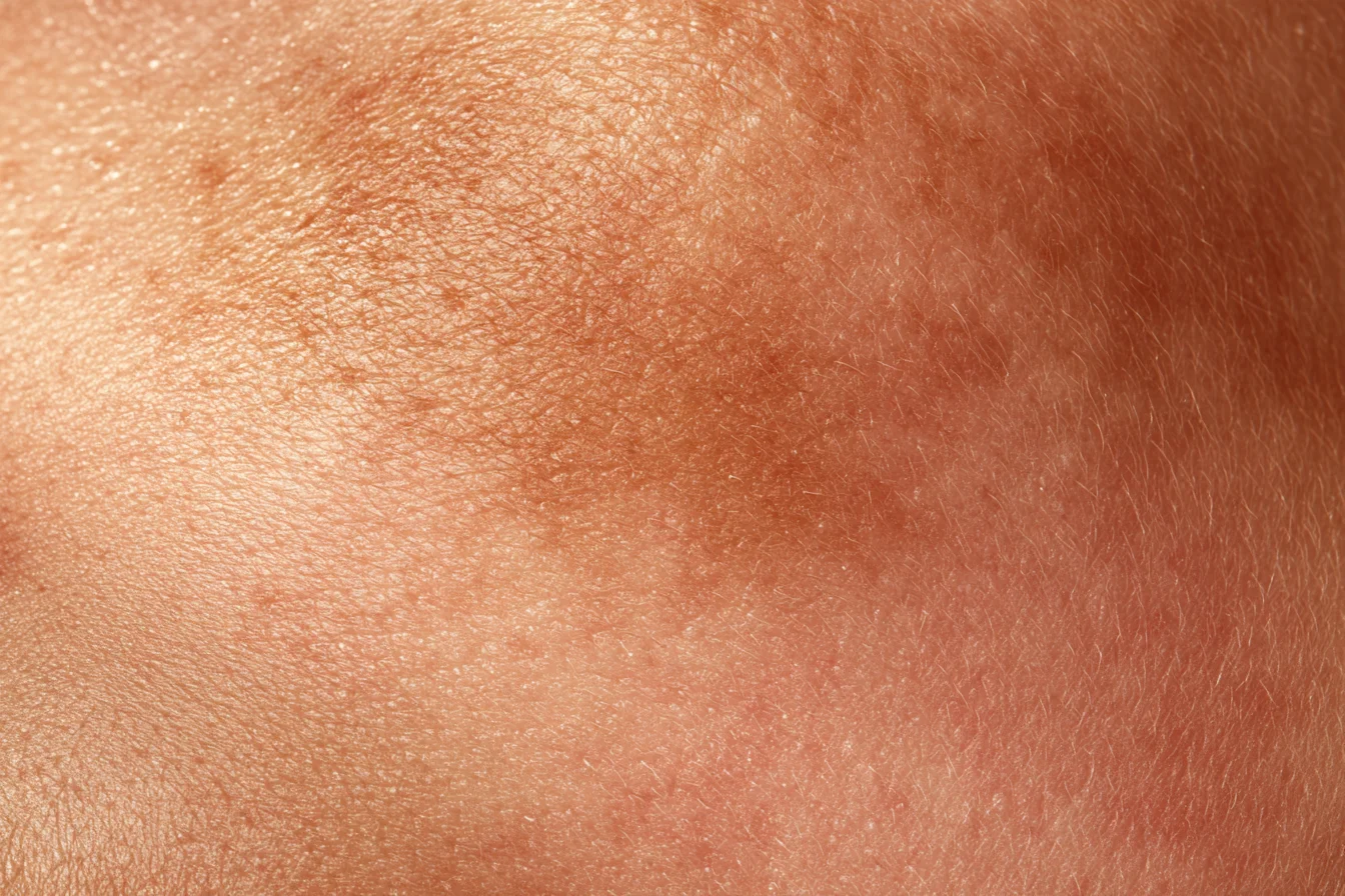
As soon as the cold wind starts, there is a remark we hear at least once in the clinic: "It had faded quite a bit last winter. But after one summer it's almost back to where it started." Up close, many of these patients have a faint red rim around the brown patches. Most have used their creams faithfully and had a fair amount of toning. Melasma usually brings to mind "brown pigment sitting on the surface of the skin", so expecting it to end once the pigment is cleared is only natural. But melasma with a red rim is a little different. Melasma is surface (epidermal) pigment layered on top of photoaging in the dermis below and an increased number of blood vessels, and the larger the dermal and vascular share, the more easily it comes back after treatments that only clear the surface pigment. That is why we plan melasma treatment by looking at the blood vessels, dermis, inflammation and barrier as well as topical medication, oral medication and toning. Quite a lot of research supporting this framework appeared in 2025–2026: a study that looked into living skin with a microscope, a randomised study that compared tissue before and after oral medication, and an international expert consensus. We go through them in turn.
Three-Line Summary
- Melasma is epidermal pigment layered on photoaging of the dermis (a broken basement membrane, solar elastosis, senescent fibroblasts) together with increased blood vessels and inflammation. The larger the dermal and vascular share, the more easily it returns after treatment that clears only the surface pigment.
- Lightening creams and oral medication did their job in reducing epidermal pigment, but in studies pendulous melanocytes, solar elastosis and vascular signals in the dermis remained unchanged within 3 months. The effect of toning alone also faded over time.
- So, on a foundation of sun protection (tinted sunscreen), we first sort out the share of epidermis, dermis, vessels and inflammation, and when redness or vessels show between the brown, we put vascular treatment such as V-Beam into the same plan. Maintenance after improvement is where the real work starts.
Why Melasma Treatment Doesn't End at Surface Pigment — Epidermis, Dermis and Vessels Layered Together
Think of a stain spreading on wallpaper. Wipe only the wallpaper (the epidermis) and it looks clean for a while. But if the wall behind it (the dermis) is damp and the pipes inside (the blood vessels) are leaking, the stain seeps through again. Melasma is just like this.
Under the microscope, melasma skin is not just a case of pigment cells (melanocytes) overworking. Several changes appear together in the dermis beneath the epidermis. These names will come up throughout this column, so here is a brief introduction to each.
- Basement membrane — the thin floor between the epidermis and dermis; think of it as the floor that keeps epidermal pigment from leaking down.
- Solar elastosis — clumped, tangled elastic fibres in dermis exposed to sun for a long time.
- Senescent fibroblasts — fibroblasts that used to repair the dermis have aged and, instead of repairing, send out signals that stimulate pigment cells.
- Increased vessels and vascular signals (VEGF, endothelin-1) — dermal vessels increase in number and size, and the vascular side releases signals that wake pigment cells.
- Mast cells — immune cells in the dermis that release inflammatory signals.
Several reviews agree that these changes closely resemble photoaged skin (Espósito 2022 review; Jo 2024 review). Simply put, melasma is a pigment problem arising on skin worn out by long sun exposure. That is why we treat melasma less as a "pigment disease" and more as "sun-tired skin signalling through pigment".
The Roots of Dermal Melasma — Photoaging and a Broken Basement Membrane
Dermal and mixed melasma are not difficult only because the pigment is deep. In melasma tissue, the basement membrane has been reported to be thinned and broken in places (Espósito 2022 review, with tissue and electron microscopy images). When the floor breaks, epidermal pigment drops into the dermis (melanin incontinence), and pigment cells may also sag down toward the dermis. Pigment cells hanging down like this are called pendulous melanocytes.
Pigment does not simply drift downward for no reason; there is also evidence that the dermis sends signals to the pigment cells above. In a tissue study comparing lesions and surrounding skin in 60 Korean women with melasma (Kang 2006), SCF — a signal that stimulates pigment cells — was increased only in the dermis of the lesions, while c-kit, which receives that signal, was increased in the epidermis. It looks as if the dermis sends the signal and the epidermal pigment cells receive it and go to work.
Senescent fibroblasts are named as one of the sources of that signal. Aged skin accumulates such senescent cells, and signalling between them and pigment cells is described as one cause of photoaging-related pigment (Kim JC 2022 review). The reason dermal melasma refills after a laser breaks up the colour is that the signalling side remains in the dermis. When our male melasma column noted that melasma in men tends to have a larger share of solar elastosis and dermal pigment, it was the same thread.
Vascular Melasma — How Increased Vessels Wake Pigment Cells
Now to the pipes. A tissue study comparing lesions and surrounding skin in 50 Korean women with melasma (Kim EH 2007) found three things. The lesions were redder than the surroundings (redness values measured with a colorimeter), and dermal vessels were increased in both number and size. Moreover, the more vessels there were, the darker the pigment, and VEGF — a signal that increases vessels — was also higher in the lesions. In short, there were more vessels under the brown patches, and more vessels meant darker patches.
Clues to how vessels wake pigment cells come from animal and cell studies. A pathway was identified in which endothelin-1 released by vascular endothelial cells drives pigment production and proliferation in pigment cells (Liu LX 2024; mice, cultured cells and cultured human skin explants). A systematic review of 34 studies on the vascular component of melasma (Masub 2020) points the same way: there are experimental and diagnostic studies showing that vascular abnormalities are involved in melasma, and the evidence for addressing the vascular component with tranexamic acid and laser or light treatments was summarised as promising.
We often meet patients like this in the clinic: press lightly on the brown melasma and release, and the redness drains first and quickly returns while the brown stays. They also say it looks darker after exercise or a sauna. For melasma like this, clearing the brown is not the end. Vascular laser can help, and we often combine vascular laser with pigment treatment. Our column on V-Beam for diffuse redness explained that vascular laser not only reduces vessels but also changes inflammatory pathways and the dermal environment, and that connection carries straight over to melasma.
For reference, melasma with increased vessels does not improve much with Skin Boosters alone. There are cases where combining Skin Boosters works well and cases that need vascular laser, so we decide on treatment by examination.
Melasma Under a Two-Photon Microscope — Where Lightening Creams Reach and What Stays
In 2025, a study looked at melasma cell by cell in living skin without a biopsy. Ning 2025 (published in JAMA Dermatology; a single-centre observational study in China; 60 patients with melasma) imaged lesions and surrounding skin in three dimensions with a non-invasive device called a two-photon microscope — a method that photographs the cell layers one at a time with light, without removing skin.
The lesions showed the following: melanin was abundant and patchy throughout the epidermis, the living epidermal layer was thinner, and the undulation of the epidermal–dermal junction (rete ridges) was flattened. At the junction there were more activated and pendulous melanocytes, and solar elastosis was more severe.
The interesting part came next. Fifty-three of these patients applied 2% hydroquinone cream for 12 weeks. The melasma score (mMASI) fell by an average of 29.81%, and melanin and activated pigment cells in each epidermal layer decreased (Ning 2025). Yet pendulous melanocytes and solar elastosis did not change statistically over the 12 weeks. The lightening cream did its job in the epidermis, but the dermal changes stayed — you can almost see on screen why melasma that faded with cream comes back when the cream stops.
That is also why we suggest using strong lightening creams such as triple combination cream in short, focused courses and moving to other ingredients for maintenance: what this cream does well is on the epidermal side. Treat the pigment itself first, and after that, maintenance treatment that strengthens the skin's foundation is needed.
Oral Tranexamic Acid for Melasma — Why It Reduces Epidermal Pigment, Yet the Dermis and Vessels Are Planned Separately
Melasma treatment today cannot be discussed without oral medication. The evidence that tranexamic acid acts on the vascular side comes mainly from animal and cell studies. In mice with UV-induced pigmentation, oral tranexamic acid reduced pigment and lowered pigment production by reducing endothelin-1 in vascular endothelial cells (Liu LX 2024; mice, cultured cells and cultured human skin explants). It was also reported that the reduction in cells carrying the vascular marker CD31 moved together with the lightening effect.
So what happened in human skin tissue? A randomised double-blind study of 50 Brazilian women with melasma (Cruz 2026) looked at exactly that. Participants were divided into tranexamic acid alone and tranexamic acid plus ketotifen (a mast cell stabiliser), took them for 3 months, and tissue from the same site was taken before treatment and on day 90 for comparison. Epidermal melanin decreased in both groups, with no difference between them — tissue confirmation that oral medication reduces epidermal pigment. However, VEGF and mast cell counts in the upper dermis stayed the same in both groups over the 3 months. By contrast, only in the group with ketotifen added did the epidermis thicken and solar elastosis, pendulous melanocytes and basement membrane breaks decrease.
We read these results this way. Oral medication clearly does its job of reducing epidermal pigment. But within 3 months it did not reach the vascular signals in the dermis, and the dermis and basement membrane changed when the inflammatory cells were addressed as well. Ketotifen is not an established melasma drug; it was tested within this study.
Our tranexamic acid column introduced tranexamic acid as suppressing vascular signals such as VEGF and endothelin, but that comes mainly from animal and cell studies. Since VEGF stayed unchanged in human tissue at 3 months, we think melasma with a large vascular component should not be left to oral medication alone, and the vascular side should be planned separately.
There are safety data as well. In a systematic review of randomised studies of oral tranexamic acid (Kuceki 2025), no thrombotic events were observed, and the 2026 global consensus (Passeron 2026) also recommends oral tranexamic acid for people at low risk of clots. Before prescribing oral tranexamic acid, we first check for contraindications such as a thromboembolic history or oral contraceptive use. We also ask about hormonal medication and smoking.
The Role of Laser Toning in Melasma — Strong on Epidermal Pigment, Not Enough on Its Own as the Vascular and Dermal Share Grows
Toning is a fundamental of melasma treatment. Low-fluence 1064 nm laser toning is the most widely used laser for melasma in Asia, and a systematic review of 42 studies (Lee YS 2022) summarised it as a generally effective and safe treatment compared with existing treatments. That the mainstay of melasma toning is still 1064 nm nanosecond was covered in detail in our nano vs pico toning column.
But melasma with a large vascular and dermal share responds differently to toning alone. In a randomised split-face study (Kaminaka 2017), 22 Asian patients with melasma or solar lentigines received 10 toning sessions on one cheek only. Melanin and redness values decreased overall, but the redness, vessels and mast cell activity that had been increased in melasma did not improve in the tissue. The researchers concluded that lesion thickness, vessels and mast cell activity are markers that help predict the effect of toning — melasma with more vessels and inflammation responds less to toning alone.
A meta-analysis of 50 toning studies with 1,772 people (Cervantes 2023) points the same way. The effect of toning alone faded over time, with almost no difference from before treatment at time points after 24 weeks of treatment, and higher effects were reported when combined with other treatments. Side effects such as white spots (hypopigmentation) were also reported more often with more sessions.
Toning works best not "a lot and often" but "as much as needed, together with other treatments". So rather than fixing the number of toning sessions in advance, we adjust the interval and number according to the response, and when vessels are visible we put vascular treatment into the same plan.
Vascular Melasma, V-Beam and Skin Botox — Evidence for the Side That Helps It Last Through Summer
Does treating the vessels as well really mean less recurrence? The first study to answer this was Passeron 2011, a randomised single-blind split-face study of 17 white women. Starting at the end of winter, triple combination cream was applied to the whole face for 4 months, and V-Beam (595 nm pulsed dye laser) was added to one side only, 3 times. When they were seen again after the summer (at least 2 months after the last treatment), the side with V-Beam had kept its improvement over baseline, while the cream-only side no longer differed meaningfully from baseline. The side where the vessels were treated as well held up through summer — this is why we put vascular treatment into the same plan for vascular melasma.
The evidence for Skin Botox (Botox placed very thinly into the dermis) has grown by one study too. In a randomised split-face study in which 28 Thai women with mixed melasma completed the protocol (Chaijaras 2025), both sides received triple combination cream for 12 weeks, and Skin Botox was added to one side only, at the start and at week 12. At week 24, three months after stopping the cream, the cream-only side was 21.03% worse than baseline, while the side with Skin Botox remained 36.97% improved. The gap that opened after the cream was stopped is the meaningful result for recurrence.
However, this study measured melasma scores, and the idea that Botox works by reducing vascular signals (VEGF) or inflammation is still an inference from animal and cell work. So, as in our Botox and pigment column, we use Skin Botox not as a substitute for vascular laser but as an option to consider together for melasma that comes with redness and a feeling of heat. Skin Boosters are sometimes added as well.
The 2026 Global Consensus on Melasma — Treating Not Only Pigment Cells but Their Surroundings
How did experts around the world sum it up? The 2026 global consensus (Passeron 2026, JEADV), produced by 10 pigmentary-disorder experts from Africa, Asia, Europe and the Americas through a literature review and the Delphi method, put a sentence in its conclusion: to manage pigmentary disorders well, not only pigment cells but the surrounding skin environment — fibroblasts, mast cells and vascular components — must be addressed. We think the key words are "not only pigment cells". Rather than putting skin-environment improvement first with Skin Boosters or other devices, the basis is still to deal with the pigment cells.
The points worth noting on melasma were these.
- Sun protection is the foundation of prevention, treatment and maintenance of melasma, and tinted sunscreen that also blocks long-wave UVA and visible light was named a good choice. The tinted sunscreen we have long been recommending is now in the recommendations.
- Oral medication and procedures are not recommended as first-line treatment for mild melasma; they have a place in moderate or more severe cases.
- Oral tranexamic acid is recommended for people at low risk of clots, and lasers and microneedling should be used very cautiously so as not to make things worse.
- After improvement, maintenance treatment to prevent recurrence is essential, and hydroquinone triple combination cream should be avoided for maintenance.
Maintenance is less about handling the surface pigment and more about addressing the broken "factory" beneath it. So for maintenance we tend to recommend Skin Boosters together with sun protection (tinted sunscreen).
Another international Delphi consensus led by a pigmentary disorders society (Sarkar 2026; 38 dermatologists from 11 countries, including Korea) reserved lasers for cases that do not respond well. This is a caution against pushing hard with lasers first, in the same direction as "don't end it with one round of toning". The 2026 answer is not one particular laser but a plan that looks at the surroundings of the pigment cells as well.
How We Plan Melasma Treatment in the Clinic — First, the Share of Epidermis, Dermis and Vessels
In the end, melasma treatment starts with knowing which side carries the larger share in your melasma.
| Layer | What is seen | Supporting evidence | What we look at together in the clinic |
|---|---|---|---|
| Epidermis | Melanin throughout the epidermis, activated pigment cells | In vivo imaging observational study (Ning 2025); biopsies in a randomised study (Cruz 2026) | Topical medication (short and focused), toning, oral medication |
| Basement membrane and dermis | Broken basement membrane, pendulous melanocytes, solar elastosis, senescent fibroblasts, dermal SCF | Tissue study (Kang 2006); reviews (Espósito 2022, Kim JC 2022); in vivo imaging (Ning 2025) | Reducing irritation, soothing and repair care, sun protection |
| Vessels | Increased vessel number and size, VEGF, endothelin-1 | Tissue study (Kim EH 2007); animal and cell (Liu LX 2024); split-face study (Passeron 2011) | V-Beam, Skin Botox, oral medication |
| Inflammation and barrier | Mast cells, skin that reddens easily with irritation | Biopsies in a randomised study (Cruz 2026); split-face study (Kaminaka 2017) | Soothing care, barrier care, no rubbing |
- Sun protection — we recommend tinted sunscreen that also blocks visible light as the basis. It applies to every row.
- Topical medication — strong lightening creams such as triple combination cream are used in short, focused courses, moving on to maintenance ingredients once things improve.
- Oral medication — tranexamic acid after checking clot risk, with the duration set by progress.
- Toning — with generous intervals, adjusted to the response rather than piling up sessions.
- The vascular side — when redness or increased vessels show between the brown, V-Beam goes into the same plan, and when heat and redness come together, Skin Botox is considered as well.
- Dermis, inflammation and barrier — we recommend soothing care such as LDM together with soothing and repair home care. Think of it as calming pigment cells that have become sensitive from irritation.
This combination and order is not from a head-to-head study but our clinical judgement based on the evidence above. Melasma needs treatment that clears it and treatment that keeps it from coming back, together. That is why improvement is where the real work begins. In particular, we recommend checking again in late spring before summer to see whether the vascular and dermal sides are rising again.
In Short — Down to the Damp Walls and the Pipes
Melasma is not a stain on the wallpaper but a skin condition that comes back less only when the damp inside the wall and the pipes are fixed too. If yours went back to square one last summer, we suggest checking the vascular and dermal sides together this winter.
Things to Keep in Mind
- The share of epidermis, dermis and vessels and the response differ between people, so results and recurrence vary even with the same treatment.
- Very frequent toning can increase side effects such as white spots (hypopigmentation).
- Vascular lasers such as V-Beam carry a risk of pigmentation depending on skin type, so we first check for redness and vessels before deciding.
- Before prescribing oral tranexamic acid, we check for a thromboembolic history, hormonal medication such as oral contraceptives, and smoking.
- Adding ketotifen was tested within a study and is not an established melasma drug. The explanation that Botox reduces vascular signals or inflammation is still an inference at the animal and cell level.
References
Show 17 references
- Passeron T, Desai SR, Abdallah M, et al. Global consensus on the management of melanin hyperpigmentation disorders. J Eur Acad Dermatol Venereol. 2026;40(5):760-772. doi:10.1111/jdv.70185
- Sarkar R, et al. Delphi consensus on melasma management by international experts and pigmentary disorders society. J Eur Acad Dermatol Venereol. 2026;40(4):680-692. doi:10.1111/jdv.70066
- Ning X, et al. In Vivo Evaluation of Melasma Pathologic Features and Treatment Response by 2-Photon Microscopy. JAMA Dermatol. 2025;161(10):1039-1047. doi:10.1001/jamadermatol.2025.2790
- Cruz ACL, et al. Histopathological evaluation of facial melasma treated with oral tranexamic acid alone and in combination with ketotifen. An Bras Dermatol. 2026;101(3):501336. doi:10.1016/j.abd.2026.501336
- Liu LX, et al. Tranexamic Acid Ameliorates Skin Hyperpigmentation by Downregulating Endothelin-1 Expression in Dermal Microvascular Endothelial Cells. Ann Dermatol. 2024;36(3):151-162. doi:10.5021/ad.23.108
- Kuceki G, Mendez D, Garza-Dueñas GG, et al. No thromboembolic events observed in randomized controlled trials of oral tranexamic acid: A systematic review and descriptive synthesis. J Am Acad Dermatol. 2025;93(4):1086-1088. doi:10.1016/j.jaad.2025.06.001
- Kim EH, Kim YC, Lee ES, Kang HY. The vascular characteristics of melasma. J Dermatol Sci. 2007;46(2):111-116. doi:10.1016/j.jdermsci.2007.01.009
- Kang HY, et al. The dermal stem cell factor and c-kit are overexpressed in melasma. Br J Dermatol. 2006;154(6):1094-1099. doi:10.1111/j.1365-2133.2006.07179.x
- Masub N, et al. The Vascular Component of Melasma: A Systematic Review of Laboratory, Diagnostic, and Therapeutic Evidence. Dermatol Surg. 2020;46(12):1642-1650. doi:10.1097/DSS.0000000000002770
- Kim JC, et al. Skin-Aging Pigmentation: Who Is the Real Enemy? Cells. 2022;11(16):2541. doi:10.3390/cells11162541
- Espósito ACC, et al. Update on Melasma-Part I: Pathogenesis. Dermatol Ther (Heidelb). 2022;12(9):1967-1988. doi:10.1007/s13555-022-00779-x
- Jo JY, et al. Update on Melasma Treatments. Ann Dermatol. 2024;36(3):125-134. doi:10.5021/ad.23.133
- Lee YS, et al. The Low-Fluence Q-Switched Nd:YAG Laser Treatment for Melasma: A Systematic Review. Medicina (Kaunas). 2022;58(7):936. doi:10.3390/medicina58070936
- Cervantes J, et al. Efficacy and Safety of Low Fluence Nd:YAG Laser Treatment in Melasma: A Meta-Analysis and Systematic Review. Dermatol Surg. 2023;49(1):36-41. doi:10.1097/DSS.0000000000003635
- Kaminaka C, et al. The Clinical and Histological Effect of a Low-Fluence Q-Switched 1,064-nm Neodymium:Yttrium-Aluminum-Garnet Laser for the Treatment of Melasma and Solar Lentigenes in Asians: Prospective, Randomized, and Split-Face Comparative Study. Dermatol Surg. 2017;43(9):1120-1133. doi:10.1097/DSS.0000000000001120
- Passeron T, Fontas E, Kang HY, Bahadoran P, Lacour JP, Ortonne JP. Melasma treatment with pulsed-dye laser and triple combination cream: a prospective, randomized, single-blind, split-face study. Arch Dermatol. 2011;147(9):1106-1108. doi:10.1001/archdermatol.2011.255
- Chaijaras S, et al. Efficacy of Botulinum Toxin A for the Management of Melasma: A Split-Face, Randomized Control Study. J Cosmet Dermatol. 2025;24(8):e70376. doi:10.1111/jocd.70376
This column is adapted from Dr. Jeong's blog post (2026-11-15, in Korean). Read the original (Korean)
From Consultation Through Treatment
A board-certified dermatologist examines you directly, identifies which layer the cause sits in, sets the device and the parameters, and then carries out the procedure as the same physician.
The number of sessions, the intervals, the maintenance point and the cost are agreed together before the first session.
Frequently Asked Questions
- Can I tell at home whether my melasma is the vascular type?
- There are hints. If the brown has a red rim, if the redness drains first when you press and release, or if it looks darker after exercise, a sauna or drinking, a larger vascular share can be suspected. But the exact share should be confirmed in the clinic with a Wood's lamp and dermoscopy.
- Is it all right to take tranexamic acid for a long time?
- In a review pooling randomised studies, no thrombotic events were observed. Still, it depends on clot history, hormonal medication and smoking, so we always check before prescribing and set the duration according to progress.
- How many toning sessions until my melasma is gone?
- It is hard to set by number. An analysis found the effect of toning alone fades over time, and toning too often can increase side effects such as white spots. We recommend adjusting the interval to the response and combining it with other treatments.
- Does everyone with melasma need a vascular laser such as V-Beam?
- No. We plan it for melasma where a vascular component has been confirmed. Depending on skin type there is also a risk of pigmentation, so we first check for redness and vessels before deciding.
- Once my melasma has faded, can I take a break from sunscreen?
- That is the most important time. The 2026 global consensus also says maintenance treatment must continue after improvement, and its foundation is sun protection. Please keep using a tinted sunscreen that also blocks visible light.
This article is general information and does not replace an individual diagnosis or treatment. Because the right decision depends on your symptoms and skin condition, please see a board-certified dermatologist.